


Pupil - the hole in the eye that lets light in
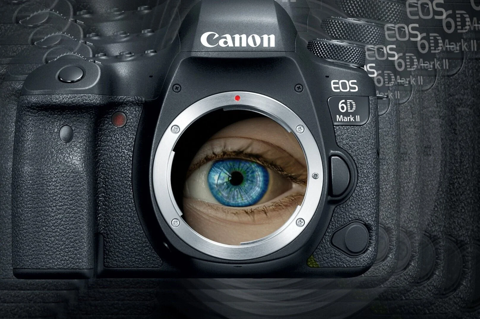
Wouldn’t it be terrible if your eye had a hole in it? But, wait a minute – it does! That hole is called your pupil, the black part of your eye in the very center. Why is it black? That’s simple – there is nothing there, just a hole which allows light from whatever you are looking at to enter your eye. Of course that hole in your eye is protected by your cornea, the clear outermost layer which covers your whole eye. Your eye is totally worthless without the pupil; you would be living in complete darkness without it. Most of us, however, never give our pupils a second thought or recognize their importance, as they do their work silently behind the scenes. A camera lens has something similar to the pupil called the aperture. The aperture is the hole that lets light in to go to the back of the camera where the light sensors are. Let’s take a quick look at how the camera aperture works.
Overexposed - too much light hitting sensors

Underexposed - too little light hitting sensors
We’ll start by figuring out what went wrong with this picture. As you can see, it’s too bright. Why? Too much light was allowed in by the aperture causing this picture to be overexposed. There are several other camera settings which might also be the culprits (ISO and shutter speed), but let’s just focus on the aperture setting, which is called the “f-stop” in photography lingo. Here’s the opposite problem; this picture is underexposed as not enough light was allowed in through the aperture to the light sensors. When your camera is set to automatic the aperture will automatically open wider when viewing a dark scene or close smaller when viewing a scene with an abundance of light. How does it know to do this? Cameras are equipped with a light meter which consists of a light sensitive material such as selenium that captures incoming light and converts this light energy into electrical energy (voltage). The more light entering, the higher the voltage, and vice-a-versa. This voltage is then analyzed and a signal is sent to small motors which adjust the blades of the aperture to open or close it.

Blades which change aperture size. Smaller numbers = larger opening
As you can see there are several very essential components involved in automatically adjusting the aperture to get the right exposure: the aperture which is composed of blades; the motors which move the blades; the light meter which measures incoming light; and a built-in software which analyzes voltage and initiates the motors to move the blades. This technology is present in even the most inexpensive cameras which are automatic.
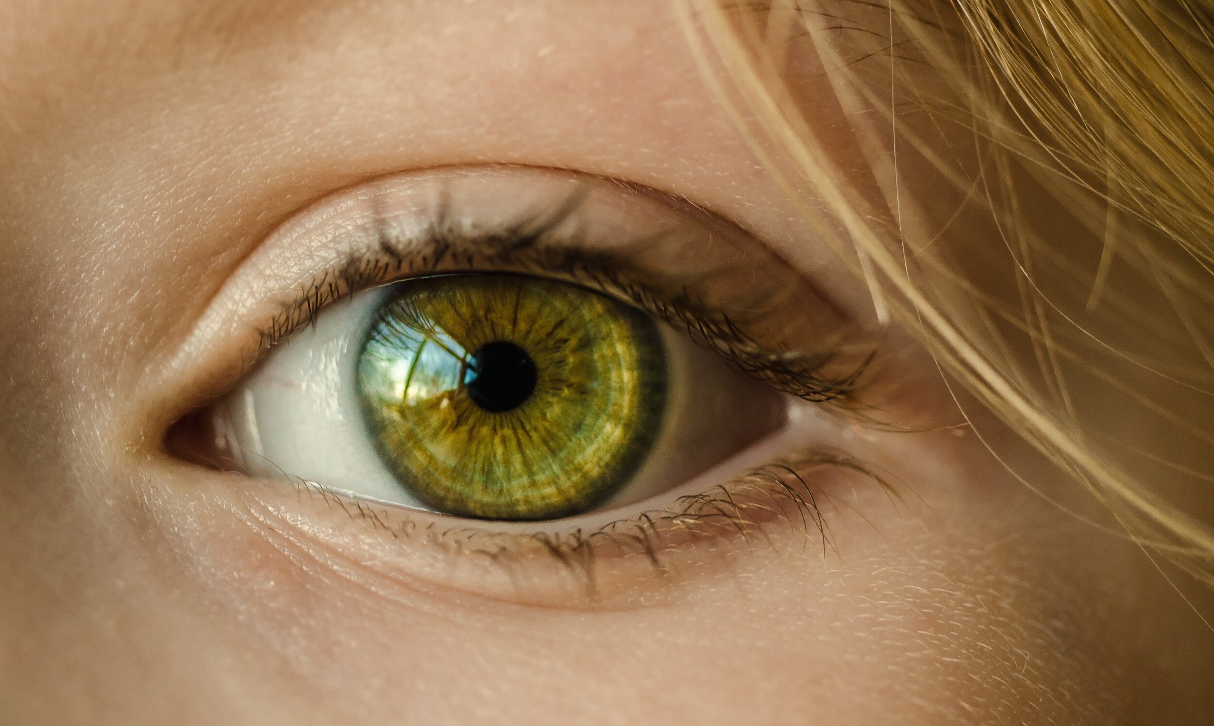
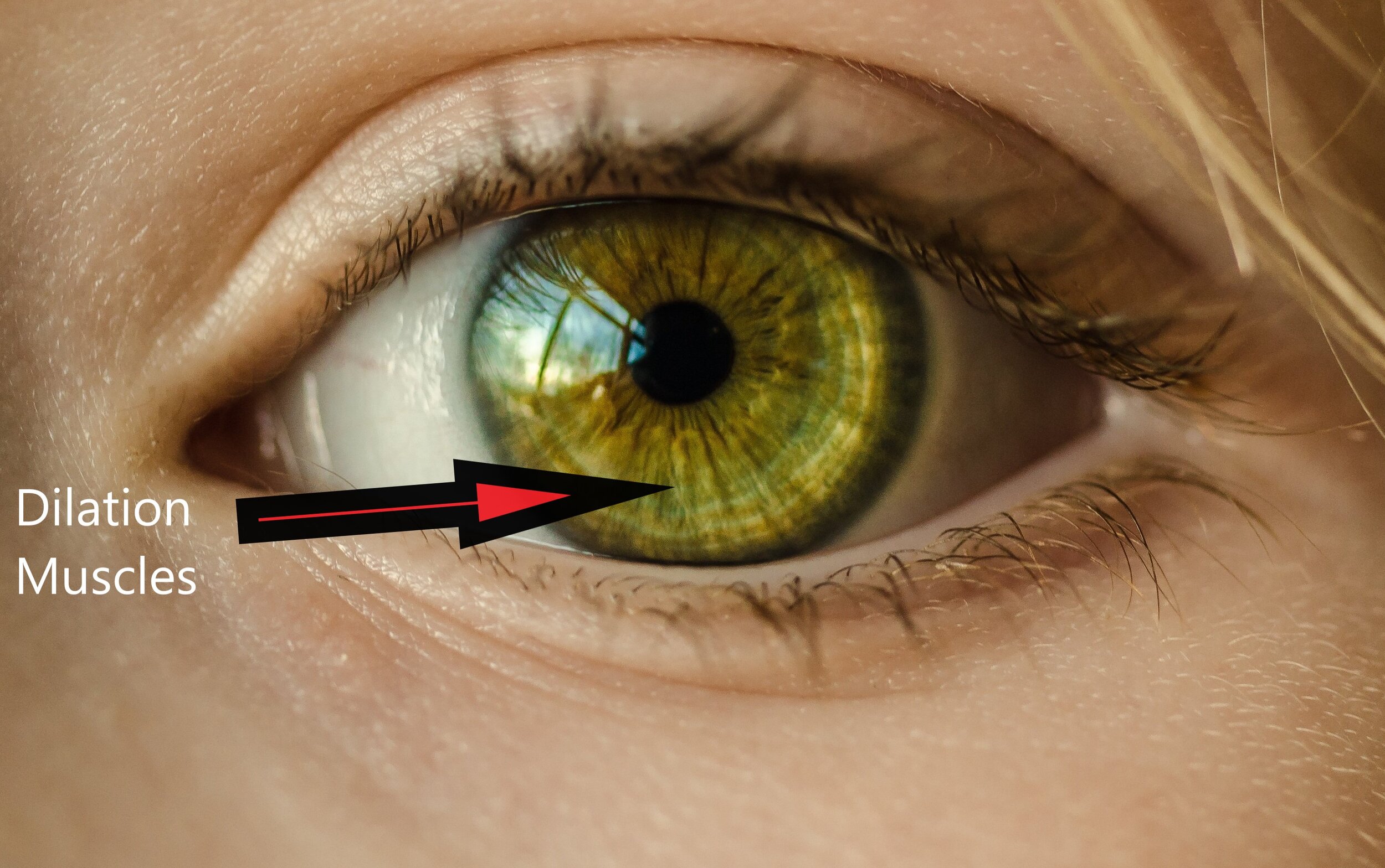
Let’s refocus on the eye. The most striking part of the eye is the colored ring around the pupil called the iris. The iris is not just there for looks, though. It plays an important role in pupillary light reflex, PLR. Hiding within its colors are two different sets of smooth muscles. Smooth muscles are not controlled by us as are our arm muscles (muscles attached to bones called skeletal muscles) , but are specially designed with automatic controls which we are totally unaware of. Smooth muscles are what move the food you’ve eaten through your digestive system. (when was the last time you consciously directed food to move through your stomach and intestines?) These smooth muscles in your iris are like the blades in the camera aperture. Let’s see how they work.


Constriction
You just walked out of a matinee showing of Star Wars into some bright sunlight. “Wow, sure is bright out here!” you say as you squint and put your hand over your eyes. But just seconds later you have forgotten all about the brightness. What happened? Without you even being aware of it your pupils constricted, meaning they closed down. Most pupils are designed to range in size from 2mm to 8 mm in diameter. When leaving the dark theater your pupils were still adjusted to very low levels of light. They were opened wide (8 mm). When you entered the bright sunlight all of a sudden an intense amount of light entered your eye. At the back of your eye are millions of rods and cones, light sensitive cells whose job it is to receive light and pass that along to the optic center of your brain which puts these messages into a colored picture of what you are looking at. However, some of these light messages veer off from the optic nerve on a separate pathway to your midbrain which sits above your brain stem. They arrive at a small group of cells there named the pretectal nucleus where they are analyzed. The pretectal nucleus then sends a signal through fibers running along the oculomotor nerve to another station called the Edinger-Westphal nucleus. Somehow this group of cells is able to recognize by the intensity of the message that the light entering the eyes is too bright. We’re in danger of having our picture overexposed, or even of permanent damage to the sensitive cells in the back of our eyes. The Edinger-Westphal nucleus then responds by firing off a message towards the eyes. The first destination of this message is another group of cells called the ciliary ganglion. The ciliary ganglion then sends a signal to the circular muscle in the iris called the iris sphincter to constrict and close the pupil down. By the way, if a bright light is shone only in one eye, the message is routed to the other eye’s ciliary ganglion so that pupil constricts also. This is what a doctor is looking for when he shines the bright light in one of your eyes. (1)

Try these activities to see for yourself how your pupil can change size
Activity: 1) Place a magnifying glass in front of a mirror and place it in front of your left eye. Watch your left pupil as you close your right eye. What happens? Why does it dilate? Now shine a flashlight in one eye as you watch the other eye. Did it also grow smaller?
2) Go into a dark room with a family member and wait several minutes. Shine a flashlight into one eye while you watch what happens to pupil size. Did you see the pupil changing size?
For more information on this activity see: https://www.exploratorium.edu/snacks/pupil

Photo by Greyson Orlando
Dilation
The next time you see someone working on a difficult math problem, check out their pupils. Surprisingly, pupils dilate while we’re working on a perplexing problem, and then return to normal size upon completion. Why is this? Dilation is controlled by our sympathetic nervous system which prepares the body for emergencies (fight or flight), so maybe we view math problems as a potential threat? An easy way to see this in action is to arouse your cat’s hunting instinct. Watch their pupils quickly change from vertical slits to a round shape which almost covers the whole iris. Here’s a video in case you don’t have a cat. Cat Pupils Dilating. The more light entering the eye increases the cat’s chances for a successful hunt. Unfortunately, we humans need more than just dilated pupils to conquer math problems!
Activity: Take a magazine into a dark area like a bathroom (there has to be a small amount of light), looking quickly at the magazine. What can you see or read? Wait 2- 3 minutes and look at the magazine again. Can you see it more clearly?
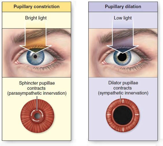
The gain in light sensitivity you experienced in those 2-3 minutes was the result of your pupils adjusting to the low light levels in the room. But, what did you do to make this happen? Not a thing! But your body has been programmed to respond to low levels of light. How? When you walk into a dark area or turn off the lights the sphincter muscle of the iris will relax, as it no longer receives messages to constrict the pupil. Your pupils will grow larger. But to really reach their maximum size and light-gathering ability the second set of muscles in your eye (dilator) need to be activated.

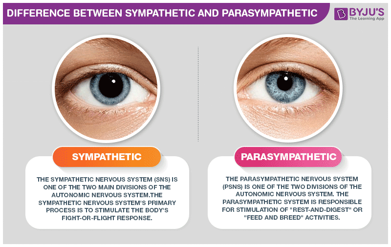
You leave your well-lit house at night to get a glimpse of the stars. At first only the brightest stars are visible, but the longer you are peering up in the darkness the more stars begin to appear. Very little light enters the eyes when you step out into the darkness. These decreasing light signals reaching the pretectal nucleus in the midbrain shut down the pupillary constriction by the sphincter muscle while at the same time activating a coordinated response through the sympathetic nervous system. I find it very interesting that a whole different branch of the nervous system controls pupil dilation than controls pupil constriction. Pupil constriction is controlled by the parasympathetic nervous system which is responsible for non-stressful activities of life like digesting food and decreasing heart rate (it is the “rest and digest” system). Pupil dilation, however, is controlled by the other part of the autonomic nervous system, the sympathetic system. This system coordinates the body’s responses to emergencies (like math problems!), things like speeding up the heart rate, and releasing adreneline and glucose into the blood stream. It is called the “fight or flight” system.

Dilation Pathway
Once the low light level message reaches the pretectal nuclues it is rerouted down from the midbrain through the brain stem to an area in the lower spine of the neck known as the ciliospinal center of budge. The cilliospinal center then initiates a response up through the neck through the superior cervical ganglion and along the carotid artery up to the ciliary nerve which then activates the dilator muscles. These are arranged like bicycle spokes radiating out from the center and when contracted (shortened) they pull the inner iris sphincter muscle outward to enlarge the pupil. (2) The same message also serves to activate the tiny muscles which elevate the eyelids, ensuring that the eye is wide open to receive any light falling on it. Full dilation can take up to 30 minutes whereas pupil constriction happens within a second or two.
There’s not a camera which comes close to our eyes in their ability to capture scenes in the detail our eyes can in the countless light environments we find ourselves in. We’ll talk more about other design features of our eyes later, but right now let’s summarize what we’ve learned about our pupils. Pupils are black because no light reflects off them; they are holes which allow light to enter our eyes. Pupils are able to automatically adjust the amount of light entering the eyes through our brain’s ability to meter incoming light and respond by controlling two different sets of muscles within our iris. This all happens without any thought or effort on our part.

Now that you know more about how your pupillary light reflex automatically adjusts the size of your pupil to bring you “pictures” with just the right amount of light exposure let’s think about what your eyes (pupils) are telling you. This automatitic and efficient system relies on a combination of important features which is, just like the camera, a result of planning and ingenuity on the part of its Designer. The One who fashioned your DNA to bring about this very intricate system has revealed Himself to you through your amazing pupils!

Our pupils allow the light reflecting off those we love to enter our eyes. This helps us understand what Moses meant when he described God’s care of His children in the wilderness: "He found him in a desert land, And in the howling waste of a wilderness; He encircled him, He cared for him, He guarded him as the pupil of His eye. Deut. 32: 10. God was keeping His children whom He loved in the “pupil of His eye”, never taking His eyes off of them. A similar phrase is used by David in Psalm 17:8, “Keep me as the apple of the eye; Hide me in the shadow of Your wings.” The phrase “apple of the eye” comes from the same Hebrew word translated “pupil” in Deuteronomy 32:10. Today, ask God for the awareness that you are in the very center of His eyes as one He loves and cherishes.
Endnotes
Yoo H, Mihaila DM. Neuroanatomy, Pupillary Light Reflexes and Pathway. [Updated 2020 Aug 10]. In: StatPearls [Internet]. Treasure Island (FL): StatPearls Publishing; 2020 Jan-. Available from: https://www.ncbi.nlm.nih.gov/books/NBK553169/
Lykstad J, Reddy V, Hanna A. Neuroanatomy, Pupillary Dilation Pathway. [Updated 2020 Aug 10]. In: StatPearls [Internet]. Treasure Island (FL): StatPearls Publishing; 2020 Jan-. Available from: https://www.ncbi.nlm.nih.gov/books/NBK535421/